
Micropatterned Slides
Slides with a micropattern for spatially defined cell adhesion for various 2D and 3D cell culture applications.
Slides with a micropattern for spatially defined cell adhesion for various 2D and 3D cell culture applications.
A micropatterned surface for cell assays with fluorescence microscopy readout.
Easy handling without preparation: Unpack and start
Excellent optical-quality imaging chamber for high-resolution microscopy
Applications
Analysis of cells using various approaches (e.g., transfection, proteomics, metabolic activity tests) with microscopy readout
Cell variability assays (e.g., CAR-T cell activity assay)
Applying defined shear stress to cell arrays (using the µ-Slide VI 0.4 product variation)
Test the attachment of your cell type on the RGD binding motif with the Trial Pack
Live cell imaging and fluorescence microscopy
Immunofluorescence staining and high-resolution fluorescence microscopy of living and fixed cell
Technical Features
µ-Slide that has a micropatterned surface with a covalently bound RGD motif (binding motif from fibronectin) on the ibidi Bioinert surface
Due to the Bioinert surface, cells can attach only on the patterned area, even during long-term cultivation for days or weeks
Bioinert surface passivation—superior to standard ultra-low attachment (ULA) surfaces:
No cell or protein adhesion
Long-term stability
Biologically inert
Bioinert is layered onto the ibidi Polymer Coverslip, which provides the highest optical quality for imaging when using high-resolution microscopy
Non-fluorescent patterns, not visible in phase contrast microscopy
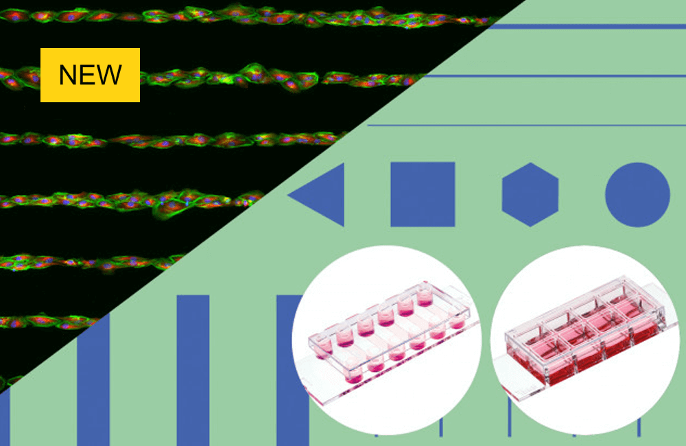
Patterns are printed on either the µ-Slide 8 Well high or µ-Slide VI 0.4
Compatible with staining and fixation solutions
Fully biocompatible materials
Also available as µ-Slides With Multi-Cell µ-Pattern and µ-Slides With Test µ-Patterns
Click here for more information